Visual Neuroscience Lab
Associate Professor
McGill Vision Research
Dept. of Ophthalmology
1650 Cedar Avenue, Rm. L7-120
Montreal, Quebec, Canada H3G 1A4
t: (514) 934-1934 ext. 36269
f: (514) 934-8216
e: janine.mendola@mcgill.ca
Binocular Rivalry from Luminance and Contrast
Although the binocular combination of luminance has been recognized for decades,
rivalry with luminance patch stimuli has rarely been examined closely. In one study
we devised a series of psychophysical studies designed to study binocular rivalry
based only on dichoptic luminance differences, and compare such results to an
equivalent series with the classically studied orthogonal gratings [Qiu et al., 2020].
Each pair of image below shows the left and right eye image. We showed that the
well-known Levelt Propositions still apply, despite the very slow (tri-stable) rivalry
we found. Our results support models of rivalry, especially those that incorporate
aspects of contrast normalization, an important computation thought to underlie
the responsiveness of visual cortex.

Localizing Rivalry Dominance and Suppression with MEG
We also designed MEG experiments of binocular rivalry that could relate to the fMRI
series described below. I believe that directly comparing the same paradigm with fMRI
and MEG might illuminate different aspects of neural coding (e.g., neural synchrony
verses magnitude of response). So far, we have used MEG when each eyes stimulus is
tagged with a different flicker frequency. This powerful technique [Bock et al., 2019].
reveals greater activity for the image that dominates awareness over a wide expanse of
visual cortex. Moreover, we discovered a new aspect of the tagged evoked response
that correlates in magnitude with individual alternation rate, reminiscent of our earlier
finding.

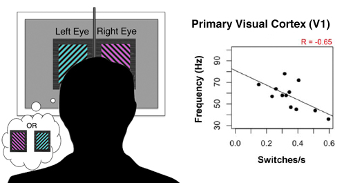
Individual differences in bistable perception in humans
As we learn more about genetic contributions to brain structures and functions, it is increasingly necessary to consider how small differences in anatomy and physiology relate to individual cognition and performance. Perceptual rivalrythe experience of alternation between two mutually exclusive interpretations of an ambiguous imageprovides powerful opportunities to study conscious awareness. It is known that individual subjects experience perceptual alternations for various types of bistable stimuli at distinct rates, and this a stable, heritable trait. Also stable and heritable is the peak frequency of induced gamma-band (30 100 Hz) oscillation of a population-level response in occipital cortex to simple visual patterns, which has been established as a neural correlate of conscious processing. Interestingly, models for rivalry alternation rate and for the frequency of population-level oscillation have both cited inhibitory connections in cortex as crucial determinants of individual differences, and yet the relationship between these two variables has not yet been investigated. We used magnetoencephalography to compare differences in alternation rate for binocular and monocular types of perceptual rivalry to differences in evoked and induced gamma-band frequency of neuromagnetic brain responses to simple nonrivalrous grating stimuli [Fesi & Mendola, 2014]. For both types of bistable images, alternation rate was inversely correlated with the peak frequency of late evoked gamma activity in primary visual cortex (200400 ms latency). Our results advance models of inhibition that account for subtle variation in normal visual cortex. Moreover, advancing our models of rivalry to the level of single subjects makes the eventual translation to subjects with abnormal binocular vision far more likely, and increases our impact further.
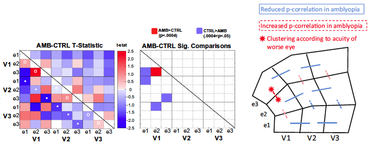
Resting State Networks in Visual Cortex
In collaboration with Dr. Amir Shmuel, I have pursued studies of functional connectivity in
networks of cortical areas activated during fMRI scanning while a participant rests (so
called resting-state fMRI), and when they complete tasks (task-based fMRI). This
method takes advantage of the full temporal resolution in fMRI time courses by
computing the pair-wise correlations between two regions of interest in order to discover
which regions are consistently co-activated and therefore probably connected [Dawson et
al., 2013]. We have looked for fine-scale retinotopic functional connections in resting
state data as a test, which we contrasted with existing independent gold standard data
on anatomical connections [Dawson et al., 2016]. Moreover, we showed recently that
adults with a history of childhood amblyopia show retinotopically specific abnormalities
in functional connections at a fine scale within primary visual cortex [Mendola et al, 2018].
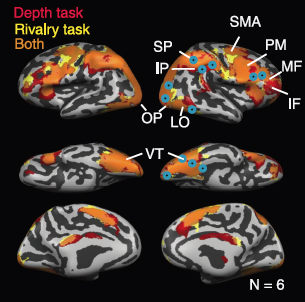
Neural substrates of depth perception and binocular rivalry in humans
The ability to perceive stereoscopic depth in the presence of rivalry has been a longstanding controversial issue with important implications for models of binocular vision. It has previously been shown that it is possible to perceive depth and rivalry simultaneously in the same location [Buckthought & Wilson, 2007]. We took advantage of these results to design an fMRI study that allowed either depth or rivalry to be reported for the same retinal stimulus [Buckthought & Mendola, 2011]. We showed that early visual areas responded equally to depth or rivalry, but dependent on the preferred spatial frequencies, providing rare physiological support for psychophysically derived channels. Results also challenged traditional theories of the correspondence problem with evidence for a mid-level representation of visual surfaces. Finally, some specific areas showed a bias for either depth (parietal cortex) or rivalry (lateral occipital, temporal cortex). We are now in a position to further characterize the responses of these regions, and make predictions regarding their response to other stimuli.
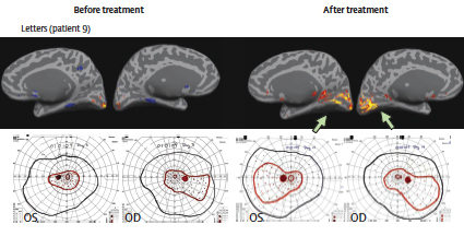
Neuroimaging Methods to Measure Treatments for Blindness, and Reorganization of Function in Visual Cortex
Given that fMRI is virtually without risk, it is safe to scan subjects repeatedly, to monitor change over time. Thus, fMRI is a natural tool for the study of longitudinal neurological treatments. Nevertheless, the number of such studies is still small, and certain issues such as test-retest reliability are not known for sure. In this collaborative project we added fMRI as an objective measure of visual function in a clinical trial of a gene replacement treatment for congenital blindness. We are the first group to scan subjects both before and after sight recovery [Koenekoop et al., 2014]. So far, the results have been encouraging. We observe perceptual gains along with increased fMRI activation post treatment. This is a unique opportunity to bridge the gap between molecular genetics and cognitive neuroscience. The work has been featured in the media, and presented at several international conferences.
Retinotopic
Organization in Children with Normal Vision
Amblyopia, commonly known as a lazy eye, is a relatively common developmental
disorder of vision. Subjects with amblyopia are characterized primarily
by poor acuity in one eye and poor depth perception. The current neurological
account of amblyopia is insufficient, and treatment has limited success.
In order to improve understanding of the neural substrates of amblyopia,
we are applying functional magnetic resonance imaging to study the brains
of children as well as adults with this condition. In a first step towards
childhood studies, we have shown, for the first time, an adult-like
pattern of retinotopic organization in children with normal vision [Conner
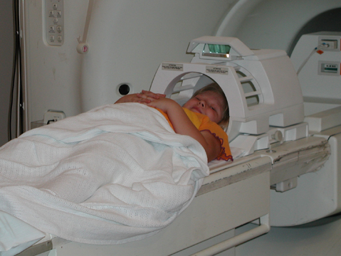
et al., 2004]. In order to improve the reliability of the young subjects,
we have installed a ‘mock’ magnet with a head motion detector
so that children can practice our experiments in a simulated environment
Anatomical
MRI of Human Amblyopia
We hypothesize that the brains of subjects with amblyopia show anatomical
and functional abnormalities, and that this information will improve
our understanding of the condition. There is evidence that the peripheral
disorders that occur during critical periods of development (monocular
blur or deviated eye) lead to sensory reorganization in the cerebral
cortex. In fact, some experts believe that in some patients central
defects in nervous system development may be causal factors that lead
to inappropriate motor control of the eyes. The long-term goal of this
project is to evaluate structural and functional anomalies of cortex
in amblyopic subjects before and during therapy as a means of evaluating
parameters that affect cortical plasticity. So far, structural MRI has
been used to identify differences in visual cortex between normal and
amblyopic children and adults [Mendola et al., 2005].
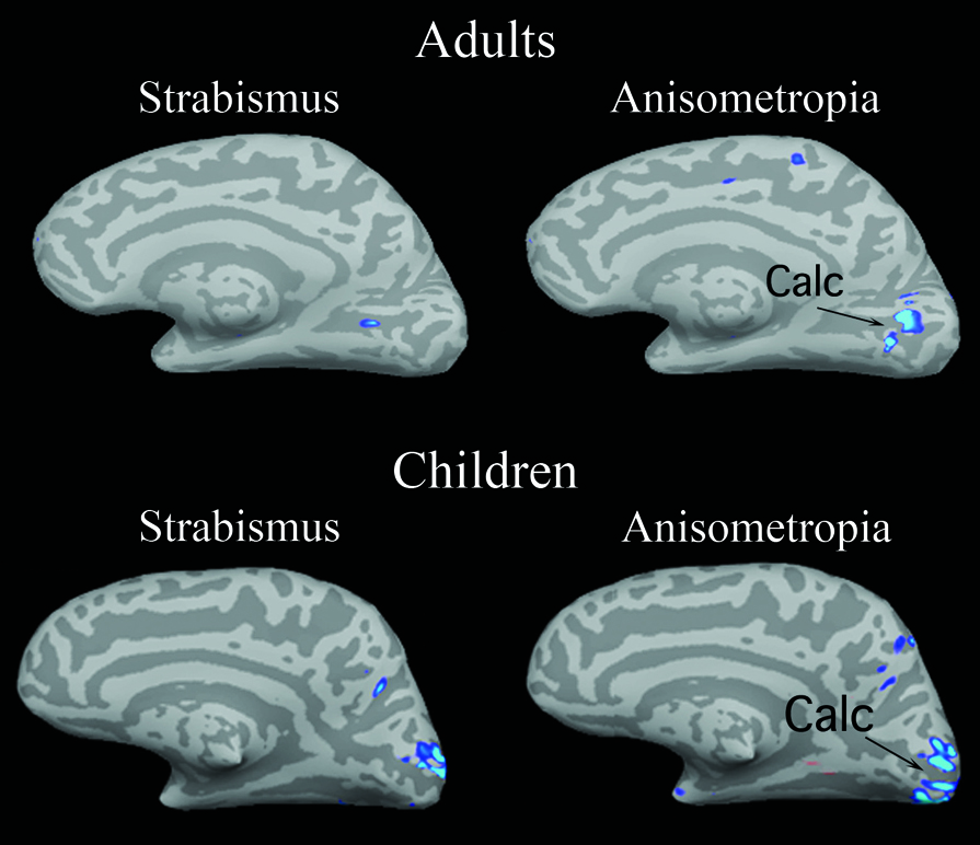
fMRI Studies of Human Amblyopia
We have obtained functional retinotopic maps from both adults and children
with anisometropic amblyopia, strabismic amblyopia, or normal vision.
The subject groups are matched for age and education within cohorts.
Our study sought to determine whether the retinotopic organization in
visual cortex mapped through amblyopic eyes are abnormal, as would be
predicted by behavioral measures. The cardinal axes of space (eccentricity
and polar angle) were mapped separately. Each eye was stimulated while
the other eye viewed an isoluminant gray screen, and fixation stability
was monitored with our Avotec-SMI system. For both adults and children,
the amblyopic eyes showed a reduced representation of the central visual
field, consistent with the well-known central visual acuity loss of
such subjects. Moreover, in certain subjects, e.g., with early onset
strabismus, we find evidence for active interocular suppression of the
central field representation of the amblyopic eye by comparing maps
when the other eye is open versus closed [Conner et al., 2007].

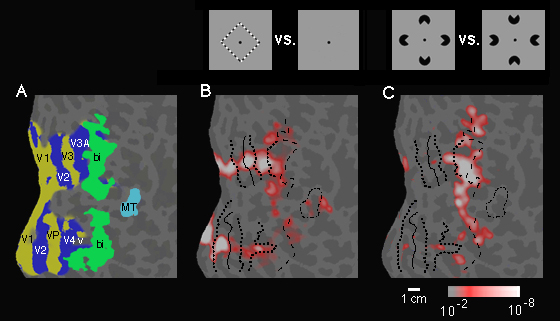
fMRI Studies of Illusory Contours
In studies of normal visual perception, often the brain is viewed as
a hard wired system of extraordinary complexity that transmits a faithful
copy of world to the organism. However, on the other hand, students
of psychology know that perception is an active, creative, and flexible
process that "goes beyond the information given." One past
experiment compared the neural representation of visible contours and
“Illusory” contours in visual cortex [Mendola et al., 1999].
We hypothesize that shapes defined by illusory contours show regions
of the brain especially important for segmentation of figures from background.
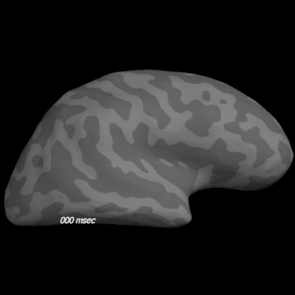
MEG Studies of Illusory Contours
In an effort to better understand the temporal dynamics of brain activity
when subjects perceive illusory contours, we have also used the Magnetoencephalography
(MEG) technique. This technique allows measure of brain activity to
be make every 5 msec. The results of this study were consistent with
our hypothesis that lateral occipital cortex is critical for the perception
of illusory shapes, and may seed ‘feedback’ information
to primary visual cortex [Halgren et al., 2003]. CLICK ON BRAIN BELOW