Localization: mapped retinotopic areas
 Image generated by Aladdin Ghostscript (device=ppmraw)
Sulci
Gyri
10
-10
V1
V1
V3
VP
VP
V1
V3
V3
VP
V2d
V2v
V2v
V2d
V2d
V2v
V4v
V4v
V4v
V3A
V3A
V3A
RA
TL
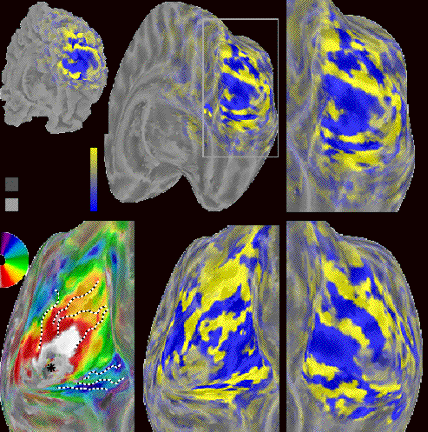
Automatic volumetric segmentation of human retinotopic cortex

€Automatic
€No cortical surface required
€Directly supplies volumes for VOI analysis
Dumoulin et al. (2003)
V1, V2, V3/VP, V4v, V3A, VO (V8)
Our specific version developed by Dumoulin, requires no cortical surface reconstruction and yields a volume of interest analysis directly. Slide with three vectors, based on rate of change: polar angle, eccentricity and aMRI.  VOI involves collapsing the voxels in each visual area, of each subject, and averaging across subjects.
Unfolded/inflated
Medial posterior
Parietal, temporal, occipital
Actual surface for each SS as marked